
On February 28, 2025, a new type of neuroendocrine tumor tracer, [18F]AlF-NOTA-LM3, developed by the Department of Nuclear Medicine at PUMCH, was officially applied in clinical practice. This new tracer demonstrates significantly superior lesion detection compared to the international gold standard [68Ga]Ga-DOTATATE, due to its lower physiological uptake in various abdominal organs, which substantially enhances the visibility of lesions. This research was carried out by Professor Huo Li's team from the Department of Nuclear Medicine at PUMCH and the result was published in the internationally prestigious European Journal of Nuclear Medicine and Molecular Imaging (IF=8.6, a tier 1 journal, or ranked among the top 5% by the Chinese Academy of Sciences).
Traditional imaging methods such as CT and MRI often fail to detect microscopic tumor lesions. To address this limitation, the new tracer employs a "molecular targeting" technique that "labels" neuroendocrine tumor cells, making them clearly visible in imaging studies. According to Director Huo, this technology demonstrates significantly higher detection rates for millimeter-sized metastatic lesions in the liver, lymph nodes, and other sites when compared to the international gold standard, achieving sensitivity of above 95% and specificity of 98%. During clinical trials, the technology successfully identified liver metastases smaller than 5 millimeters—sub-centimeter lesions that would typically remain undetected by traditional techniques.
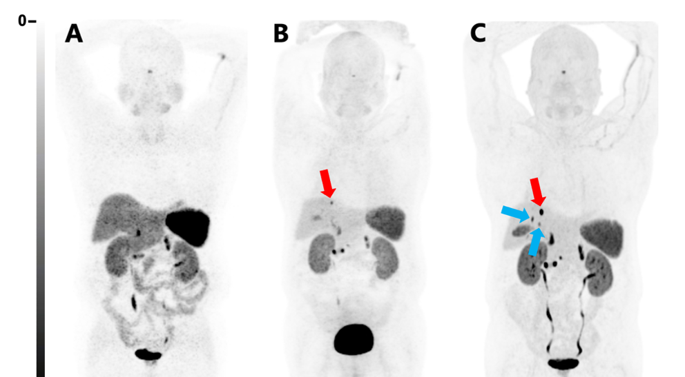
▲[18F]AlF-NOTA-LM3 detecting metastatic lesions, confirming its extremely high sensitivity
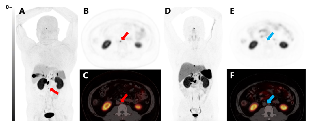
▲[18F]AlF-NOTA-LM3 detecting metastatic lymph nodes smaller than 5 millimeters that [68Ga]Ga-DOTATATE failed to identify
The ability to detect sub-centimeter microscopic lesions represents a huge breakthrough for precise staging and individualized treatment of neuroendocrine tumors and other conditions. Director Huo emphasized that this tracer offers multiple clinical applications: it can precisely locate primary lesions, provide comprehensive whole-body staging, and monitor treatment efficacy. Beyond neuroendocrine tumors, the tracer is applicable for other conditions with high somatostatin receptor expression, including pheochromocytoma/paraganglioma and tumor-associated hypophosphatemic osteomalacia. For patients with suspected tumor-associated hypophosphatemic osteomalacia, it rapidly identifies pathogenic lesions; for neuroendocrine tumor patients, it helps screen candidates suitable for targeted radiotherapy, enabling precise targeting of tumor cells while preserving normal tissues.
In comparison to imported alternatives, this newly implemented tracer is marked by higher production yields and a longer half-life, which facilitates transportation to multiple medical centers. The Department of Nuclear Medicine at PUMCH has been conducting PET-CT examinations using somatostatin receptor antagonists since 2018, developing substantial expertise in this field. With more translational research on somatostatin receptor antagonists than any other medical institution globally, PUMCH has demonstrated both the safety and efficacy of its related patents through clinical validation in over 300 cases.
Dr. Huo Li stated that the Department of Nuclear Medicine at PUMCH remains committed to pursuing innovation in tumor diagnosis and treatment, having evolved from “follower” to “leader” in nuclear medicine technology development. The department is keen to implement more innovative technologies that will benefit cancer patients and improve their prognosis and quality of life.
Written by Li Congxin, Dong Jingge, Hong Chenwei, and Chen Xiao
Pictures courtesy of Li Congxin and Dong Jingge
Edited by Dong Jingge