A 28-year-old man had a tumor that grew in his lung, extended to his heart, and then was washed down in the bloodstream to his brain, kidney, intestines and coronary arteries. The repeated episodes of tumor embolism made chemotherapy impossible, and the surgery was extremely difficult because the tumor extensively invaded the heart and lungs. Recently, the team of Miao Qi, Director of the Department of Cardiac Surgery of PUMCH, worked with other departments to successfully remove the heart and lung tumors completely for the patient and preserve as much healthy lung lobe area as possible through in situ transplantation of autologous lung lobe, creating favorable conditions for his postoperative recovery.
At the end of 2021, the patient Mr. Liang suddenly developed symptoms of cough and fever, which were not relived by pneumonia-related treatment. Seeing a huge opacity in the right lower lung on the lung CT, his doctor realized he might have a lung tumor, but such diagnosis could not be established after repeated needle biopsies. Just when the doctor was still considering how to take a sample for pathological analysis, Liang had sudden abdominal pain, and the enhanced CT showed that the tumor had invaded the left atrium and then dislodged, causing vascular blockage in the left kidney and intestines. At his wit’s end, that local doctor suggested Liang take his chances in Beijing.
Considering Liang was very young and his family were adamant for getting the best possible treatment, Miao’s team, after several rounds of evaluations, concluded that the surgery could defuse the ticking “bomb” that could go off any minute in his heart, and could also create favorable conditions for subsequent comprehensive treatment and shed light on the pathological diagnosis.
On the day of hospitalization, Liang experienced chest tightness, chest pain and temporary memory loss simply because of some increased activities in the preoperative examination. The examination results confirmed that the tumor had detached again and clogged the blood supply vessels of the heart and brain. Upon learning about that, the cardiac surgery team did not waste a moment and rushed to finish the preoperative evaluation and preparations, while organizing multidisciplinary discussions to formulate treatment plan and contingency plans for various situations that might occur during the operation with input from the Department of Anesthesiology, Intensive Care Unit (ICU), Oncology, Cardiology, Respiratory, and Thoracic Surgery.
Intraoperative exploration revealed that the tumor in Liang’s lung had invaded the left atrium along the pulmonary vein, as well as the important surrounding structures such as the pulmonary hilum, chest wall and diaphragm. The cardiac surgery team removed the heart tumor and the whole right lung. For the sake of Liang’s post-operative recovery and chemotherapy, the team painstakingly stripped the right upper lung lobe from the eviscerated right lung that was invaded by the tumor and re-implanted it back into the chest cavity after proper trimming of the pulmonary vessels and bronchi.
The surgery took 6 hours. After surgery, Liang’s transplanted lung functioned well and recovered rapidly, and he was discharged from the hospital 10 days later. Currently, he had successfully received the first session of chemotherapy and immunotherapy in the Department of Respiratory.
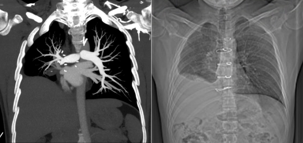
Two months after surgery, imaging showed normal blood flow and ventilation in the patient’s autologously transplanted right lung lobe, with normal activity tolerance and lung function.
Correspondent: Liu Jianzhou and Zhao Yanxue
Reporter: Hong Chengwei
Picture courtesy: The Department of Cardiac Surgery
Translator: Liu Haiyan
Editor: Liu Xingrong and Wang Yao