The 55-year-old Mr. Huang underwent a dangerous and rare surgery, the resection of a rare giant paraganglioma in the right retroperitoneum, in Peking Union Medical College Hospital. The multidisciplinary team of PUMCH innovatively applied combined thoracoscopic and laparoscopic surgery and removed the mass successfully.
In 2019, Mr. Huang started to have a headache without inducement, sometimes dozens of times a day. Strangely enough, his headache eased when he was standing. During the headache attack, Mr. Huang's blood pressure also increased to 220/130 mmHg. In March 2020, Mr. Huang's symptoms worsened, seriously affecting his work and life. Once again, he was examined and found having a huge tumor hidden in his body. It was a paraganglioma with an incidence rate of only 1/100000. Mr. Huang began to seek medical treatment everywhere, and had went to many large hospitals, but no surgery was performed. With one last hope, he came to PUMCH.
Professor Li Hanzhong from the Department of Urology did intensive research on the location and anatomical structure of the tumor. The tumor grew on the right side of the abdominal aorta at the level of the T10-L1 vertebral body, behind the inferior vena cava, with a maximum length of 9cm. Part of the tumor was in the thoracic cavity, about 3 cm × 4 cm; and the rest was in the abdominal cavity, about 5 cm × 4 cm. Compared to most of paragangliomas, Mr.Huang’s tumor located higher, crossing the thoracic and abdominal cavity. Thoracoscopic or laparoscopic surgery alone was unable to completely remove the tumor, which increased the difficulty of the operation. Furthermore, the tumor, with abundant blood supply, was close to the spinal nerves, major blood vessels, and liver. How to completely resect the giant paraganglioma without damaging the spinal nerves and avoiding hemorrhage was the key of the surgery.
Prof. Li attached great importance to this case. Before the surgery, the Office of Medical Affairs organized a multi-disciplinary consultation. Experts from the Departments of Endocrinology, Vascular Surgery, Orthopedics, Thoracic Surgery, Hepatic Surgery, Anesthesiology and Critical Care Medicine attended the meeting and made a detailed surgical plan together.
At 11 a.m. on January 11, 2021, a combined operation of urology and thoracic surgery began. Surgeons creatively applied thoracoscopic and laparoscopic surgery at the same time. The two sets of equipment were separated by only a thin diaphragm. The entire exploration process was clearly and vividly showed on the two monitors of the thoracoscope and laparoscope. A local protrusion of the psoas major muscle in the right phrenic angle could be seen under laparoscope, and thoracoscope showed part of the tumor clearly. The inner side of the huge tumor was closely adhered to the vertebral body and inferior vena cava. Many blood vessels penetrated into the tumor from the chest wall, abdomen wall and vertebral body. The complicated location of the tumor added enormous difficulties to the surgery. Even a minor mistake could bring serious consequences to the patient.
However, everyone's determination was not staggered. During the operation, with the cooperation of the anesthetists, the chief surgeon opened the diaphragm as planned, took a combined thoracic-abdominal incision, separated the tumor from the surrounding severely adhering tissues, ligated the related blood vessels, and finally resected the tumor completely.
After the operation, Mr. Huang’s blood pressure was stable and he recovered well. The postoperative pathological diagnosis was compound paraganglioma (paraganglioma + ganglion cell neurofibroma + some ganglion cell neuroblastoma components). This is the first reported case of compound paraganglioma wrapped by the psoas muscle in the right phrenic angle. And it is also the first case to use combined thoracic and laparoscopic surgery to remove the tumor.
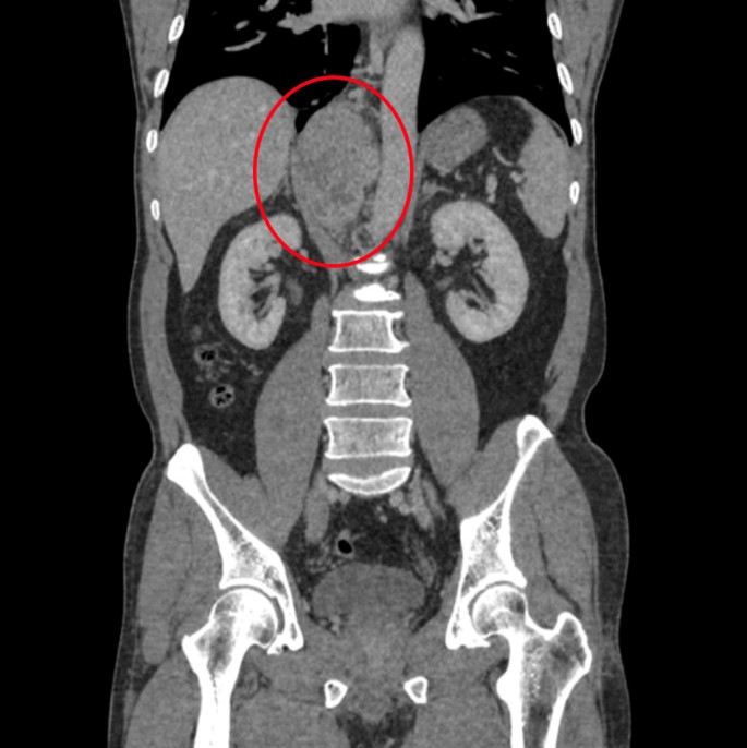
The major axis of the paraganglioma was 9cm.
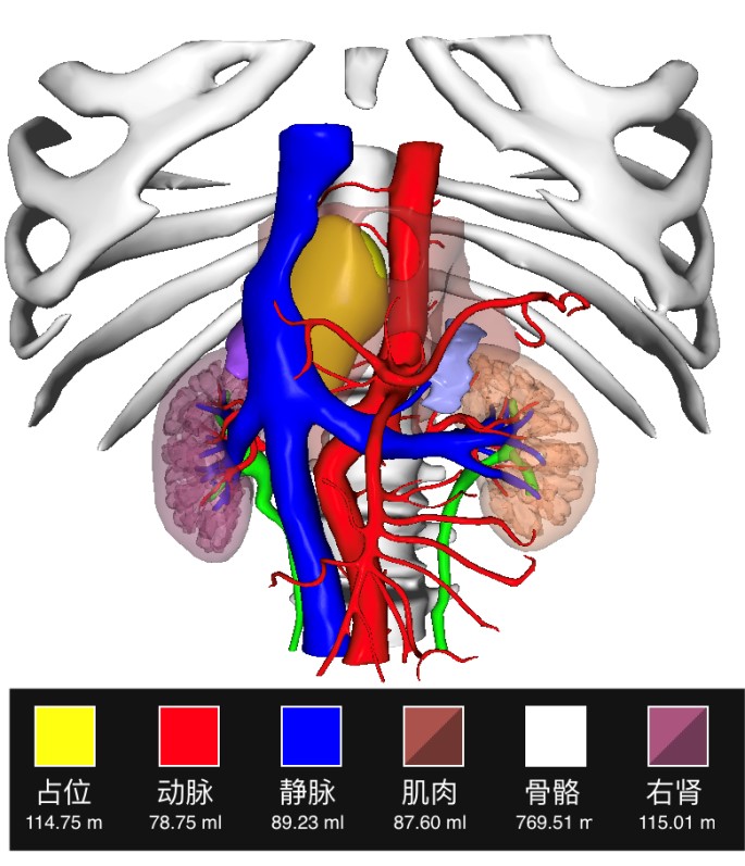
3D print showing the location of the paraganglioma
Text: Liu Chang, Wen Jin, Xu Xiaohui
Pictures: Department of Urology
Translator: Guo Chao
Editor: Kang Lin