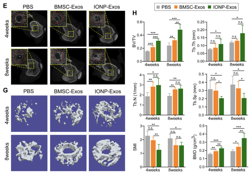
本研究开发了一种新型来源于磁驱动(氧化铁纳米颗粒(IONPs)与磁场相结合)的骨间充质干细胞外泌体(IONP-Exos)。研究结果发现,与普通干细胞外泌体相比,IONP-Exos能显著防止隧道周围骨质流失,促进更多的骨质长入肌腱移植物,增加肌腱-骨隧道界面的纤维软骨形成,并诱导更高的最大失效载荷。同时发现IONPs-Exos通过miR-21-5p/SMAD7 信号通路调控肌腱与骨整合的纤维化过程,为促进组织工程中肌腱到骨的愈合提供一种新策略。

Figure 1. (E) Micro-CT images of the cross-sections of the tibial bone tunnels at 4 weeks and 8 weeks after ACLR. (G) Reconstructed 3-dimensional models of micro-CT images of newly formed bone at 4 weeks and 8 weeks after ACLR. (H) Quantitative results of new bone formation within the bone tunnel as measured by micro- CT at 4 weeks and 8 weeks after surgery. Micro-CT parameters: bone volume fraction (BV/TV); trabecular thickness (Tb. Th); trabecular number (Tb. N); trabecular separation (Tb. Sp); structure model index (SMI); bone mineral density (BMD).
期刊杂志:Materials Today Bio
影响因子:10.761
主要作者和团队成员:吴向东、康琳、田静静、吴远浩、黄岳、刘洁颖、王海、邱贵兴、吴志宏
使用仪器:激光共聚焦显微镜
主要内容:使用激光共聚焦显微镜观察荧光标记外泌体的细胞摄取和细胞内定位
代表性 figures:

Figure 2. (E) Confocal images showed that the red fluorescence dye PKH26-labeled BMSC-Exos and IONP-Exos were endocytosed by NIH3T3 fibroblasts after 4 h, 12 h, and 24 h of incubation. F-actin was stained with phalloidin (green), and the nuclei were stained with DAPI (blue).
文章链接:https://www.sciencedirect.com/science/article/pii/S259000642200117X

