Peking Union Medical College Hospital (PUMCH), together with Peking University People’s Hospital, Wuhan Third Hospital, and Huangshi Central Hospital, among others, has achieved a major research advance in AI-assisted diagnosis of pulmonary nodules. The team developed a novel deep learning model, DeepFAN, and conducted Asia's first registered clinical trial evaluating the AI-assisted diagnostic tool for distinguishing benign from malignant pulmonary nodules. The model has received Class III medical device registration from China's National Medical Products Administration (NMPA), providing a clinical decision support tool backed by high-level evidence for intelligent, standardized diagnosis of pulmonary nodules. The findings were published in Nature Cancer, a leading international oncology journal.
Of all malignant tumors, lung cancer has the highest incidence and mortality rate in China. With the widespread use of chest CT, growing numbers of incidental pulmonary nodules are being detected, and distinguishing benign from malignant nodules efficiently and accurately has become a major challenge in radiological diagnosis.
DeepFAN innovatively integrates three key modules: a Vision Transformer that captures global features of the nodule and its surrounding microenvironment; a fine-grained 3D residual network that extracts local details such as density, lobulation, and spiculation; and a graph convolutional network that deeply fuses the two. The model was trained on 11,438 pathology-confirmed pulmonary nodules from multiple centers. Its overall ability to classify benign and malignant nodules was assessed using the area under the curve (AUC), where values closer to 1 indicate higher diagnostic accuracy; the model achieved an AUC of 0.939 on internal testing. External validation using data from the U.S. National Lung Screening Trial yielded an AUC of 0.943, with a predictive value for benign nodules as high as 0.992—supporting precise exclusion of benign nodules and helping to avoid unnecessary follow-up for patients.
The clinical trial included 463 pathology-confirmed nodules from 400 patients across three centers, with 12 junior radiologists (1 to 5 years of experience) doing two rounds of image reading. DeepFAN alone achieved a diagnostic AUC of 0.954. Without AI assistance, the readers' average AUC was only 0.667; with AI assistance, their average AUC rose to 0.776—an improvement of 10.9%, and diagnostic accuracy improved by 10.0%, sensitivity by 7.6%, and specificity by 12.6%, while interreader diagnostic consistency improved by 34.5%. Analysis showed that the model relied predominantly on global features, complementing the diagnostic approach of radiologists, who tend to focus more heavily on local features.
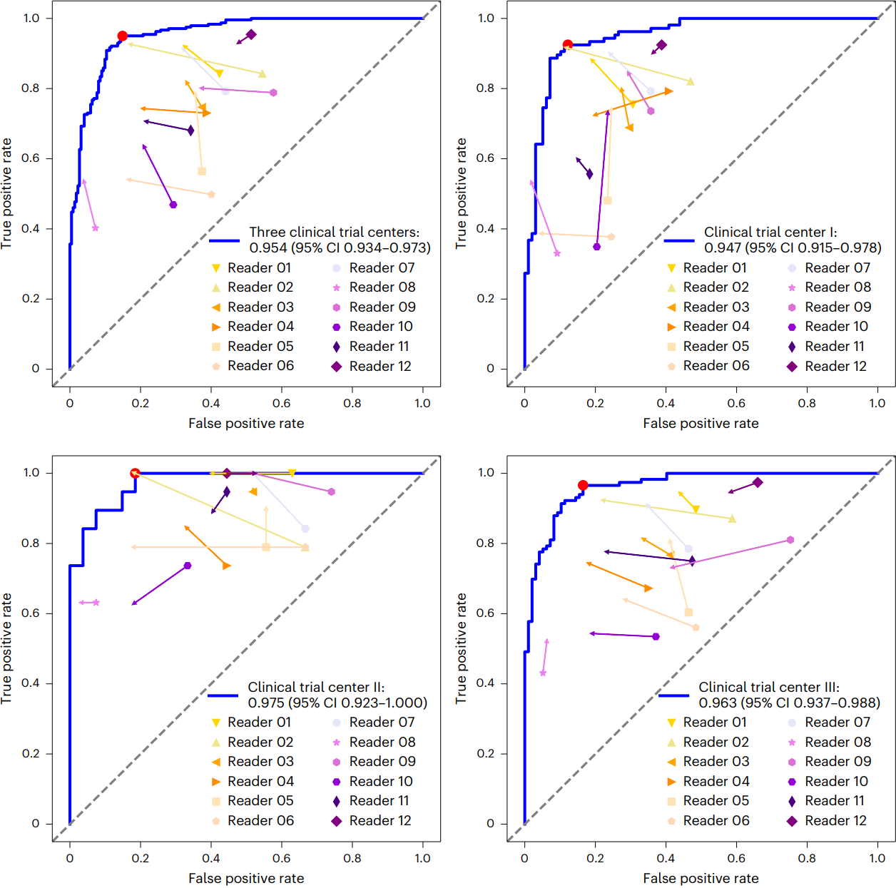
▲ROC curve for DeepFAN diagnosis and changes in diagnostic performance with AI assistance
This study provides an effective tool for the intelligent differential diagnosis of pulmonary nodules, helping to improve consistency of care across regions and setting a benchmark for research on AI-assisted standardized imaging diagnosis.
Co-corresponding authors of the paper are Song Lan, Associate Chief Physician, and Jin Zhengyu, Chief Physician, both of the Department of Radiology at PUMCH; Hong Nan, Chief Physician of the Department of Radiology, Peking University People's Hospital; and Professor Yu Yizhou of The University of Hong Kong. Co-first authors are Zhu Zhenchen, Resident Physician of the Department of Radiology at PUMCH; Hu Ge, Assistant Researcher of the Institute of Clinical Medicine, PUMCH; and Sun Chao, Senior Engineer of the Department of Radiology at Peking University People's Hospital.