
Recently, a collaborative research team led by Dr. Chen Youxin from the Department of Ophthalmology and Dr. Feng Jun from the Department of Hematology at Peking Union Medical College Hospital (PUMCH) has published a paper titled “Multimodal Imaging Characteristics and Risk Factors Analysis of Waldenström Macroglobulinemia Retinopathy” in “American Journal of Ophthalmology”. The paper offers comprehensive insights into the multimodal imaging characteristics of Waldenström macroglobulinemia retinopathy (WMR), one manifestation of the rare disease Waldenström macroglobulinemia (WM), and identified serum IgM levels and M protein levels as indicators with good predictive value for WMR. This research finding has important clinical significance for the fundus examination of WM patients.
Waldenström macroglobulinemia, also known as primary macroglobulinemia, is a type of B-cell non-Hodgkin’s lymphoma characterized by lymphoplasmacytic infiltration of the bone marrow and elevated levels of monoclonal immunoglobulin (IgM). WM can result in multi-system damage. WMR, a clinical manifestation of primary macroglobulinemia, is characterized by dilated and tortuous retinal veins, giving a “sausage link” appearance, retinal hemorrhage, and optic disc edema etc., and, in some cases, severe visual impairment. Previous studies primarily focused on individual case reports and offered limited insights into the clinical characteristics and multimodal imaging analysis, and no research on the relationship between systemic and ocular risk factors.
This retrospective, cross-sectional study included 50 patients who underwent ophthalmic examinations and were diagnosed with WM at PUMCH from 2012 to 2022. Among them, 28 patients had WMR in at least one eye. The study summarized the multimodal imaging characteristics of WMR and analyzed potential risk factors.
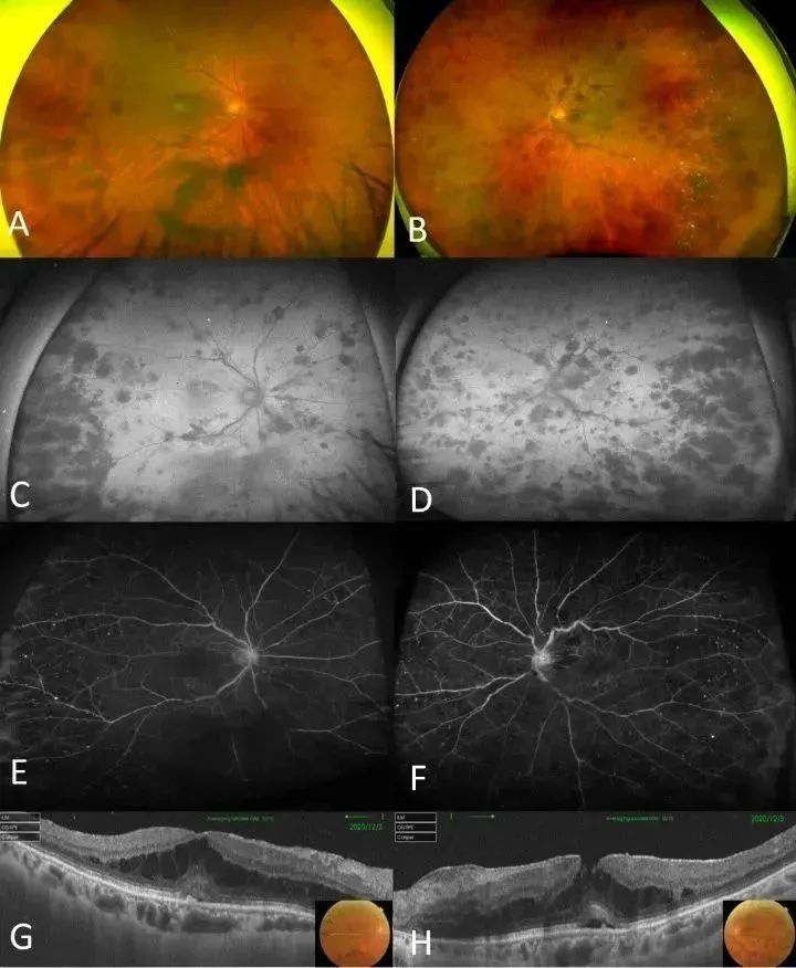
▲Multimodal images of a 74-year-old female patient with WMR
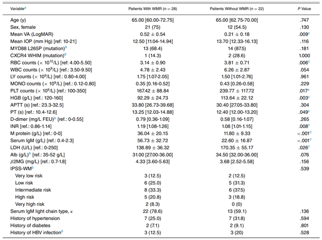
▲Clinical characteristics of WM patients with WMR
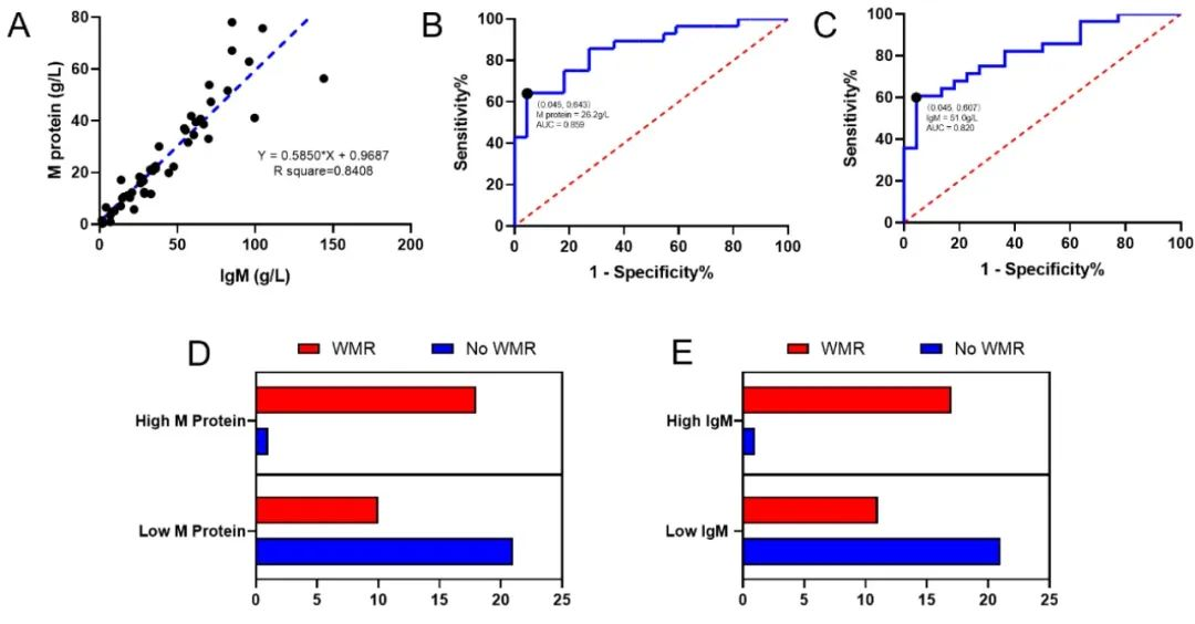
▲Analysis of correlation between serum M protein and IgM levels and WMR occurrence. The study identify the optimal cutoff values of 26.2 g/L for M protein and 51.0 g/L for IgM. By categorizing WM patients based on the cutoff values of M protein and IgM, the sensitivity for predicting WMR was as high as 95.4%
The research results reveal that WMR has distinct characteristics across various imaging modalities. UWF (ultra-widefield) laser scanning of fundus showed “sausage link” changes of retinal vessels, and UWF-AF (autofluorescence) of fundus showed extensive blockage of autofluorescence due to hemorrhage. UWF-FFA (fundus fluorescein angiography) demonstrated extensive peripheral retinal ischemia. Wide-field OCTA (optical coherence tomography angiography) of the retinal layer demonstrated noticeably tortuous vessels and the characteristic serous macular detachment. Both univariate and multivariate analyses found that serum IgM levels and M protein levels are reliable predictive indicators. The optimal cutoff value for M protein is 26.2 g/L and 51.0 g/L for IgM, which yielded a sensitivity of 95.4% and specificity of over 60% for both.
Given that WMR is a specific manifestation of WM, it is recommended that patients diagnosed with WM undergo fundus examinations to monitor their IgM or M protein levels. If these levels reach or exceeded 51.0 g/L or 26.2 g/Land fundus examination confirms the presence of WMR, immediate medical intervention is advised.
Quote
Chen H, Wang Y, Xu Z, Li D, Du H, Chen Y, Feng J. Multimodal Imaging Characteristics and Risk Factors Analysis of Waldenström Macroglobulinemia Retinopathy. Am J Ophthalmol. 2023 Mar 22; 253:233-242. doi: 10.1016/j.ajo.2023.03.011.
Edited by Wang Jingxia and Chen Xiao
Translated by Liu Haiyan
Reviewed by Zhang Xia and Wang Yao