Recently, the outcomes of project applications for the 2023 National Natural Science Foundation of China (NSFC) were announced. Two PUMCH experts, Wang Yining, Chief Physician and Deputy Director of the Department of Radiology, and Yang Meng, Chief Physician of the Department of Diagnostic Ultrasound, were granted the National Science Fund for Distinguished Young Scholars. Below is a brief introduction for us to learn about these two distinguished young scholars.
Wang Yining: Building a New Non-invasive Cardiovascular Imaging System
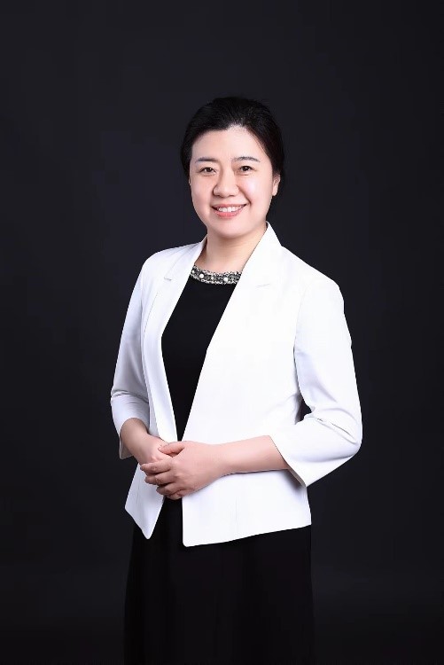
Wang Yining is Deputy Director and Chief Physician of the Department of Radiology, and also a doctoral supervisor. She has published 116 papers as the first/corresponding author in journals such as “JACC: Cardiovasc Imaging”, “ACS Applied Materials & Interfaces”, and “European Radiology”. Her research findings have been recommended by seven domestic and international guidelines and consensus. Wang Yining was granted 14 invention patents (including four international patents filed under PCT), with two already commercialized. She led 15 projects including a Major International Joint Research Project under NSFC, and a Key Project supported by the Beijing Municipal Natural Science Foundation. She has authored four published expert consensus. She was granted one prize of the National Science and Technology Progress Award and three first prizes of provincial- or ministerial-level science and technology awards.
In recent years, Wang Yining has dived into non-invasive cardiovascular precision imaging. Specifically, she conducted systemic and in-depth research on three aspects: precision quantitative assessment of myocardial injury, myocardial injury-specific multimodal molecular imaging, and new methods of contrast agent-free cardiovascular imaging using magnetic resonance. These efforts have paid off and she secured multiple innovative achievements: establishing a precision quantitative assessment system for myocardial injury, providing key evidence that informs treatment decision-making; establishing a specific diagnostic system for myocardial injury using multimodal molecular imaging, offering scientific basis for risk stratification of myocardial injury and precision and individualized diagnosis and treatment; proposing a series of new methods for contrast agent-free cardiovascular imaging using magnetic resonance, significantly improving diagnostic accuracy and safety.
The series of research achievements by Wang Yining have provided new ideas, technologies, and methods for the diagnostic imaging of cardiovascular diseases featuring “precision, specificity and safety”. These advancements have contributed to the development of the world’s first 5.0T ultra-high-field MRI, a cutting-edge equipment that China developed on its own.
Yang Meng - Overcoming Core Challenges of Photoacoustic Imaging
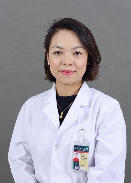
Yang Meng is the Chief Physician and Assistant to Director of the Department of Diagnostic Ultrasound, and also a doctoral supervisor. She, as the first/corresponding author, had 75 papers published in or accepted by journals such as “Radiology”, “Biomaterials”, and “Photoacoustics”. She filed for/was granted 18 invention patents (including two international patents filed under PCT), with one already commercialized. She has led 10 projects including one supported by the Key Program of NSFC, one by the General Program of NSFC, one by the Young Scientists Fund of NSFC, and one by the first Distinguished Young Scholars program of the Beijing Municipal Natural Science Foundation. Yang Meng participated in the formulation of 11 international standards. She received seven prizes, including the first prize of China Invention Innovation and Entrepreneurship, the second prize of the Ministry of Education Science and Technology Progress Award, and the first prize in the Youth Category of the Capital Medical Innovation Translation Competition.
Yang Meng has long been conducting innovative research focused on the theory and key technologies of photoacoustic imaging with the goal of expanding imaging technologies for medical functions and enhancing diagnostic capabilities based on multidimensional medical information. Yang Meng has successfully addressed the common core challenges of photoacoustic imaging, including specific identification of tissue oxygen, clear display of tissue oxygenation, and the correlation between blood oxygen and disease diagnosis. Such research efforts have significantly improved the sensitivity, resolution, and accuracy of photoacoustic imaging. The core technology was applied to the first photoacoustic-ultrasound multimodal imaging equipment in China, breaking the monopoly of imported equipment. Internationally, Yang Meng has pioneered unique functions such as real-time fusion imaging of tissue structure and oxygen saturation, as well as quantitative diagnosis of blood oxygen with both intelligent technologies and photoacoustic imaging. These advancements have expanded the clinical application scope and promoted industrial development of photoacoustic imaging.
Yang Meng’s research findings have enhanced the capabilities to collect and analyze important biological information such as human tissue structure and functional metabolism and enabled non-invasive, real-time, and quantitative in vivo imaging of tissue oxygen. These research outcomes therefore provide new means for revealing the physiological and pathological mechanisms of the human body, establishing new strategies for the diagnosis and treatment of major diseases such as tumors.
Compiled by Chen Xiao
Translated by Liu Haiyan
Reviewed by Wang Yining, Yang Meng, and Wang Yao