
Recently, the patent for “A Method of Automatic Whole-gland Prostate Segmentation Based on CNN Deep Learning” invented by Prof. Jin Zhengyu and Prof. Sun Hao’s team from the Department of Radiology, PUMCH was granted. This whole-gland prostate segmentation method, in line with the anatomical characteristics of the prostate gland, is more accurate than conventional methods in clinical practice. It can be used for automatically determining prostate cancer staging and assisting in radiotherapy planning, among others. The study received funding support from the “central high-level special program of clinical research”.
The accurate automatic whole-gland segmentation of the prostate on magnetic resonance (MR) images can significantly reduce the segmentation time and improve the segmentation consistency. This allows it to play an important role in the management of prostate cancer and benign prostate hyperplasia and demonstrate significant clinical value in the automatic determination of prostatic cancer staging and radiotherapy planning. In recent years, deep learning-based automatic segmentation techniques have been explored and applied in prostate disease imaging. However, most of the previous studies were based on single-center datasets or single sequence images, and some of the segmentation models were not able to adapt to new data, and the models failed to accurately predict heterogeneous external datasets and underperformed in generalization. Unsolved problems included that the huge variations in the resolution of prostate MR images of different sequences were not sufficiently considered, and that the segmentation difficulties were not approached from the angle of the prostate anatomical structure.
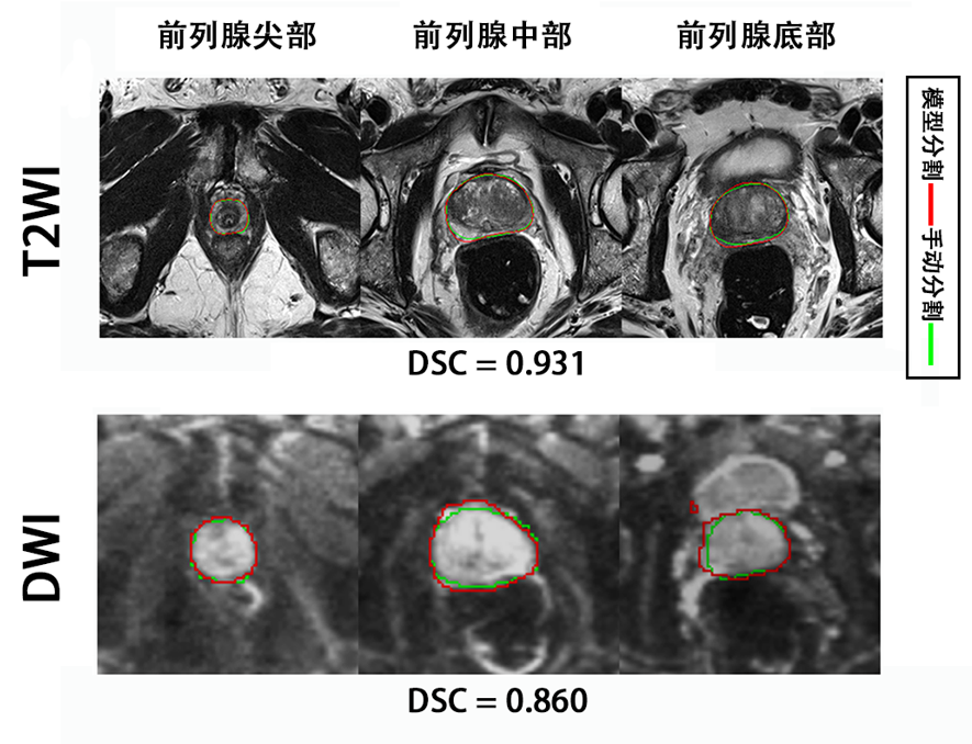
▲Example of segmentation results: On the T2WI and DWI image, the automatic segmentation (red line) and manual segmentation (green line) overlap to a large degree at the apex, mid and base portions of the prostate (from left to right), and the overall DSC of segmentation is 0.931 on the T2WI and 0.860 on the DWI.
Based on the characteristics of prostate MR images and the prostate anatomical structure, the radiology team of PUMCH established a CNN deep learning-based method, device, system and storage medium for automatic whole-gland prostate segmentation. This method divides the prostate MR images into various types such as T2WI, DWI, ADC or DCE images, and takes the apex, mid and base portions of the MR images of the prostate as inputs into the whole-gland segmentation network. Once the apex, mid and base images of the prostate are generated as outputs, the results obtained by automatic segmentation will be combined to finally arrive at the whole-gland prostate segmentation images.
By training the segmentation model for different zones of the prostate separately, the method systemically addresses the differences in resolution of images of different sequences. In addition, it made adjustments during uploading and downloading of sample modules respectively. On top of that, the team took into full account the anatomical structural differences of the prostate, trained the segmentation networks for the apex, mid and base portions of the prostate separately and went the extra mile to reinforce the model for the prostate apex segmentation, so as to improve the overall segmentation performance.
According to Prof. Sun Hao, via optimizing the training process and strengthening the weak modules, the technique achieves more accurate whole-gland prostate segmentation. Thus, it can assist in rapid outlining of radiotherapy target areas in cases of prostate cancer and enable rapid analysis of whole-gland morphological features of the prostate based on automatic segmentation.
Written by Chen Xiao
Pictures courtesy of Sun Hao
Translated by Liu Haiyan
Edited by Xiao Xiong, Zhu Liang and Wang Yao