
On February 28, Luo Yaping from the Department of Nuclear Medicine, and Yang Huaxia from the Department of Rheumatology of PUMCH made important progress in the world’s first clinical study to evaluate the activity of rheumatoid arthritis (RA) using 68Ga-FAPI, which was published in “Radiology” (IF:29.146), a top international journal of radiology.
In this study, 20 RA patients (15 women, 55±10 years old) with moderate to high disease activity in the Department of Rheumatology of PUMCH were selected for clinical and laboratory disease activity assessment, as well as 68Ga-FAPI and 18F-FDG PET/CT examinations. The results of the study showed that the accuracy of the new imaging modality of evaluating RA activity using 68Ga-FAPI as a tracer was better than that of the conventional clinical evaluation modality. Among 244 affected joints in 20 patients, PET joint counts and PET joint index scores were positively correlated with the score of RA disease activity and the degree of joint damage assessed by imaging. 24.5% higher detection rate of joint lesions by PET/CT than by physical diagnostic joint examination may provide a basis for early and accurate diagnosis of RA. 15 (6.1%) joints were not detected by 18F- FDG, but were detected in the PET/CT with 68Ga-FAPI as a tracer, and 68Ga-FAPI was slightly better than 18F-FDG as a tracer for detecting RA.
Based on this clinical trial, the research team introduced the “PET Joint Index”, which provides a more objective basis for accurate assessment and treatment of RA by quantitatively reflecting the RA activity. This study was supported by funding from the “special program of clinical research for central high-level hospitals”.
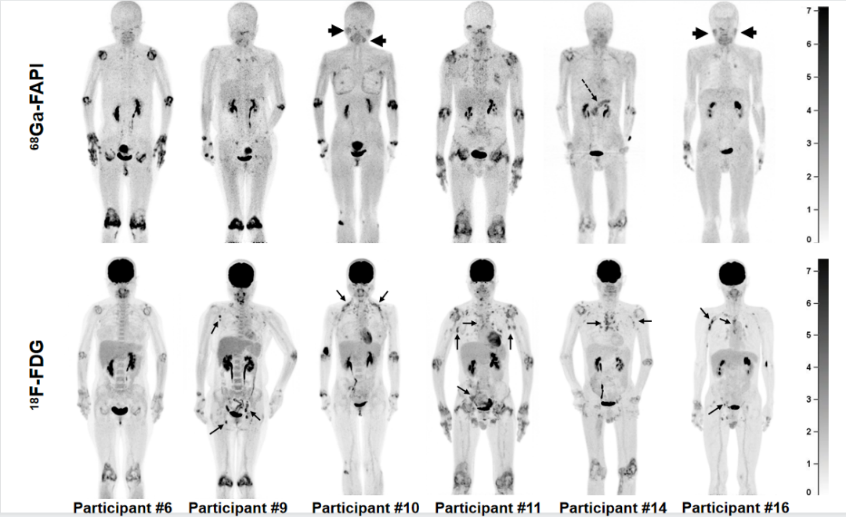
▲ Comparison of 68Ga-FAPI and 18F-FDG PET/CT maximum intensity projection image of 6 patients with RA. 68Ga-FAPI PET/CT showed more intense uptake in the affected joints
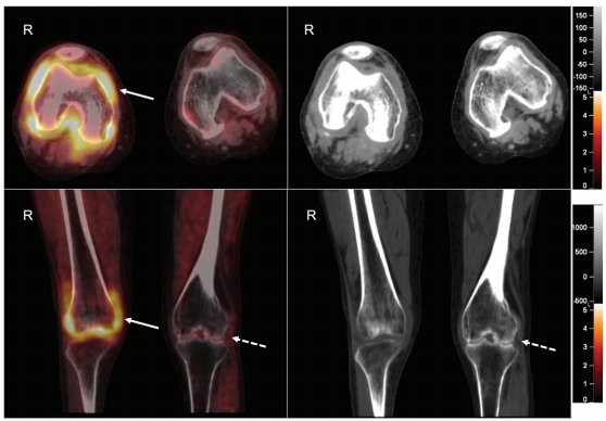
▲ The image of the knee lesion in a patient with RA showed significantly higher 68Ga-FAPI uptake and synovial thickening in the right knee joint, suggesting RA disease activity in the right knee (arrow). The left knee joint shows joint stenosis and subsurface bone destruction without 68Ga-FAPI uptake (dashed arrow), suggesting secondary osteoarthritis and non-RA disease activity in the left knee joint
Translated by Liu Haiyan
Edited by Pan Qingqing and Wang Yao