On the evening of July 1, 2020, the multidisciplinary team of Peking Union Medical College Hospital and the First Affiliated Hospital of College of Medicine, Zhejiang University (the First Hospital of Zhejiang University) collectively conducted a joint check for a patient with heart failure and severe pulmonary hypertension, discussing whether he needs a heart transplant or a heart-lung transplant.
▲The Department of
Cardiology tries to cooperate with brother hospital trough cloud, it not
only better serves patients from other places, but also actively
promote the construction of the Department of Cardiology.
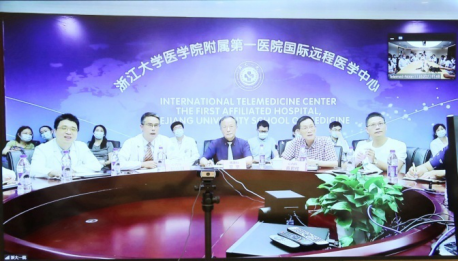
Jointly medical check with a cloud connection
The First Hospital of Zhejiang University reported an abnormal case of heart failure with severe pulmonary hypertension. The patient was a 60-year-old male with chest tightness and shortness of breath for 9 months. The electrocardiogram showed a myocardial infarction, but the coronary angiography results were normal. Recently, his heart failure continued to deteriorate and pulmonary hypertension repeatedly became worse, which made medical treatment be throny. What is the diagnosis of this patient? How should he be treated now? should a pure heart transplant by applied? Or should a combined heart-lung transplantation be considered? The two centers built a team of elite experts to solve the problems.
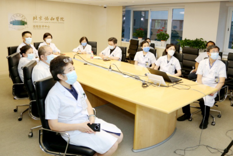
Secretary Zhang Shuyang of PUMCH attached great attention to this case and participated in person. Experts such as Zhu Wenling, Yan Xiaowei, Tian Zhuang, Xu Xiqi, Guo Xiaoxiao from the Department of Cardiology, as well as Professor Liang Zhiyong from the Department of Pathology, Professor Huo Li from the Department of Nuclear Medicine, and Professor Wang Yining from the Department of Radiology were all present. A lively discussion with the expert team of the First Hospital of Zhejiang University was conducted through video connection.
▲The Department of Cardiology of PUMCH and the First Hospital of Zhejiang University conducted remote group consultation.
Precise diagnosis and treatment of severe cases
The patient’s condition, radiographic data, and pathological pictures under the electron microscope were clearly displayed one by one through video. Each expert provideed his/her basis from their own professional fields for a clear diagnosis. Professor Zhang Shuyang keenly pointed out that the patient’s image and electron microscope are in line with the characteristics of myocarditis, and there is a possibility of viral infection. At the same time, attention should be paid to the patient’s immune system involvement. He suggested a further identification of the virus strain and an active antiviral treatment, which pointed out the direction for diagnosis and treatment. Associate Professor Tian Zhuang believed that a large number of mitral valve regurgitation should be considered as papillary muscle insufficiency rather than primary valve disease, and that the abnormal upper ventricular wall motion didn’t accord with the distribution of coronary blood supply, adding more evidence for the diagnosis of myocarditis. Director Huo Li of the Department of Nuclear Medicine pointed out that neither the myocardial perfusion nor metabolic defects of nuclide meet the coronary blood supply area, combined with normal coronary angiography, suggesting a non-ischemic cardiomyopathy. Wang Yining, deputy director of the Radiology Department, pointed out that the myocardium had diffuse edema and extensive fibrosis, and the subepicardial involvement was more prominent. The cardiac MRI findings had reached the 2009 Lake Louise myocarditis diagnostic criteria. Director Liang Zhiyong of the Department of Pathology concluded that under the electron microscope, not only the necrosis of myocardial cells and the rupture of muscle fibers were seen, but also a large number of homogeneous particles with a diameter of 20-25nm were seen inside and outside the mitochondria, suspected to be virus particles.
So far, the discussion had turned white-hot, everyone is astonished at this unprecedented change under the electron microscope. As a cardiologist, Associate Professor Xu Xiqi, also assistant to the director of the Department of Cardiology, reported the clinical and pathological characteristics of myocarditis in time, and proposed that the pathological diagnosis of myocarditis should also improve immunohistochemistry, and immunosuppression and antiviral therapy could be considered in the next step. Yan Xiaowei, deputy director of the Department of Internal Medicine, rationally pointed out the unique pathology of the patient's myocardial tissue, that the infiltration of inflammatory cells is not prominent, and the particle diameter of 20-25nm is much smaller than that of conventional virus particles such as COVID-19.
Professor Wenling Zhu agreed to arrange an elective surgery of heart transplant. Director Jing Zhicheng proposed that the small particles of uniform size under the electron microscope needed to be further studied by molecular etiology experts. Additionally, the pulmonary artery pressure fluctuated with treatment and left heart function, suggesting that it was not chronic pulmonary vascular disease, but rather pulmonary hypertension. Due to insufficient evidence of pulmonary vascular involvement, heart transplantation could be performed alone without a combined heart-lung transplant.
Photo by/Wang Pengfei