
Recently, PUMCH Liver Surgery, supported by many other departments, successfully removed a 20cm liver tumor for a 42 year old woman. The surgery not only removed the abdominal pain which troubled her for years, but unburdened her frame which is rather short due to scoliosis.
The patient was accepted by PUMCH on October 18. For years she has been suffering abdominal pains, which intensified in recent two months. Ultrasound in a local hospital indicated a giant space-occupying lesion and CT showed a tumor about 20 cm in diameter, which extended from liver to pelvic cavity and pressed other organs. The tumor was suspected to come from liver. When she was young, the patient suffered severe scoliosis, which was only partly corrected by surgery and her height remained at 154cm. In the recent two months her conditions worsened. She had abdominal pains and malnutrition, was unable to lie flat and found it difficult to walk. Her weight dropped by 7kg. The patient, with a big belly in her frail frame, sitting in wheelchair, finally landed at PUMCH after being turned down by several hospitals.
Liver Surgery assessed the case: there will be no life quality without surgery, and the tumor may rupture at any time to threaten life, while a surgery will be highly risky. The department had detailed discussions on perioperative care, operative approach and emergency response. Professor Zhong Shouxian made supplements according to his own experience. Doctors from other departments, namely, Radiology, Anaesthesiology, Operation Room, Blood Transfusion and ICU, were also invited for consultations.
The day before surgery, Professor Pan Jie from Radiology had abdominal angiography for the patient, made sure that the blood supply was from hepatic artery and conducted super-selective embolization. The day of surgery, Doctor Gong Yahong from Anaesthesiology watched over monitoring measures and vascular access. Professor Mao Yilei and Associate Professor Du Shunda from Liver Surgery performed the operation. Upon opening the abdomen one could see tissue edema and ascites. The liver looked a light red, smooth and soft. A giant tumor grew downwards out of the V, VI sections, about 20cm ×20cm×15cm, partly grey and partly red; it was of a hard texture, pressing against the abdominal side in the right, pushing the left liver to anterior axillary line in the left and its lower part entering the pelvic cavity. The gallbladder was pressed flat, omentum, transverse colon, stomach and some other parts were closely attached to the tumor and its back was unreachable. After a comprehensive survey, the surgeons performed according to plan, and removed the tumor, part of liver, part of invaded transverse colon and gallbladder successfully. Less than 500ml blood was lost, and the tumor weighed 3.5kg. To cope with hemodynamic changes due to changes in abdominal pressure, the patient spend the transition time in ICU. Then the patient’s symptoms were reduced, and she was able to walk. After ten days, the patient was discharged.
(Caption)
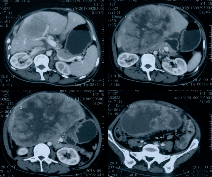
CT indicated that the tumor grows out of liver, and runs downward into pelvic cavity, pushing adjacent tissues out of their places.
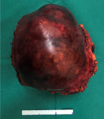
Surgery scene and the tumor removed